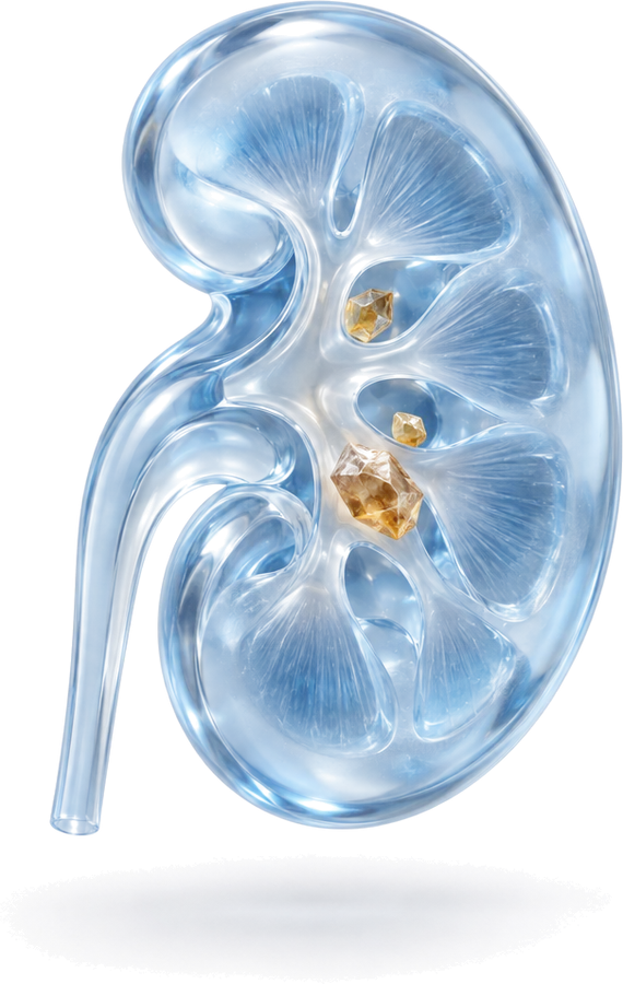
What is ureteropelvic junction (UPJ) obstruction?
Urine produced in the kidney collects in a funnel-shaped space in the middle of the kidney (the renal pelvis) and from there passes down to the bladder through the ureter. The point where the renal pelvis joins the ureter is called the ureteropelvic junction (UPJ).
In UPJ obstruction, this junction does not allow urine to flow freely. Urine builds up in the renal pelvis and the kidney’s collecting system becomes dilated; this is called hydronephrosis. Long-standing, significant obstruction can cause the kidney tissue to lose function over time. UPJ obstruction is one of the most common causes of kidney dilatation in children; it can also occur in adults.
Causes: congenital and acquired obstruction
Congenital obstruction is the most common type. The section of ureter at the junction does not have a normal muscle structure and cannot push urine forward; sometimes the ureter joins the renal pelvis at a higher point than it should, which also impairs drainage. Another common cause in adults is a blood vessel running to the lower part of the kidney and crossing in front of the ureter (a lower pole vessel), which presses on the junction.
Acquired obstruction can develop after stones, previous surgery or endoscopic procedures in this area, inflammatory processes and, rarely, tumours.
Symptoms
In babies and children
Today, a large proportion of UPJ obstructions are detected when dilatation of the baby’s kidney is seen on a prenatal (antenatal) ultrasound. Most of these babies have no symptoms at birth. Older children may have abdominal or flank pain, episodes of vomiting, urinary tract infection, blood in the urine or a palpable swelling in the abdomen.
In adults
- Flank pain that increases especially after drinking plenty of fluids, tea, coffee or alcohol
- Recurrent urinary tract infections, kidney infections with fever
- Kidney stone formation
- Blood in the urine
In some adults, the obstruction causes no symptoms at all and is found by chance on an ultrasound performed for another reason.
How is it diagnosed?
The aim is to establish whether the dilatation is really due to an obstruction and how much the kidney is affected.
- Ultrasound: Involves no radiation; it shows the degree of dilatation of the renal pelvis and calyces and the thickness of the kidney tissue. In babies with prenatally detected dilatation, the first ultrasound is usually performed a few days after birth, because the baby’s urine output is low in the first 48 hours and the dilatation may appear smaller than it is.
- Diuretic renogram (MAG3 or DTPA scintigraphy): The key test for diagnosis and the decision to operate. It shows how much each kidney contributes to total function (split renal function) and how quickly the kidney drains after a diuretic is given.
- Computed tomography (CT) or MR urography: Useful, especially in adults, for detailed assessment of the anatomy and for detecting a crossing vessel and accompanying stones.
- Voiding cystourethrogram: May be requested in children if the dilatation could be caused by backflow of urine from the bladder to the kidney (vesicoureteral reflux).
- Blood and urine tests: Kidney function and the presence of infection are assessed.
The process for babies with prenatally detected dilatation
Seeing dilatation of the baby’s kidney on a pregnancy ultrasound can be worrying for families; however, in most of these babies the dilatation is harmless and decreases over time. The usual approach after birth is as follows:
- The course of the dilatation is monitored with ultrasound during pregnancy; in most cases, the timing or method of delivery does not change because of it.
- In the first days after birth (usually after 48 hours), the baby’s kidneys are reassessed with ultrasound.
- Depending on the degree of dilatation, a second ultrasound is planned a few weeks later; if considered necessary, a diuretic renogram and voiding cystourethrogram are requested.
- In some babies, preventive antibiotics may be recommended to prevent urinary tract infection.
- Babies with mild dilatation and preserved kidney function are monitored with ultrasound at set intervals.
During this period, if the baby has fever, irritability, vomiting or feeding difficulties, a doctor should be consulted without delay to check for a urinary tract infection.
When is surgery needed?
Not every patient with hydronephrosis needs surgery. Especially in babies, a large proportion of dilatations decrease over time or remain stable without harming the kidney; these children are monitored with regular ultrasound and, where needed, a renogram. In children, the decision between monitoring and surgery is made together with the family within a pediatric urology approach.
Surgery is usually considered in the following situations:
- Function of the affected kidney on the renogram is significantly low or decreases during follow-up
- Drainage on the renogram is significantly impaired and dilatation increases during monitoring
- Presence of symptoms such as flank pain
- Recurrent infection or kidney infection with fever
- Kidney stone formation accompanying the obstruction
The decision is made individually, assessing all these findings together with the examination and test results.
Treatment: laparoscopic pyeloplasty
Dismembered (Anderson-Hynes) pyeloplasty
The standard surgical treatment for UPJ obstruction is pyeloplasty. In dismembered (Anderson-Hynes) pyeloplasty, the most widely used technique:
- The narrowed junction segment is removed completely.
- If necessary, the excessively enlarged renal pelvis is reduced in size and reshaped.
- The end of the ureter is opened to create a wide opening and is stitched without tension to the lowest point of the renal pelvis.
- If there is a crossing vessel, it is preserved and the new connection is placed in front of it.
If there are kidney stones together with the obstruction, they can be removed during the same operation in suitable patients.
Laparoscopic (keyhole) approach
In suitable patients, pyeloplasty is performed using the laparoscopic method, with a camera and fine instruments inserted through several small openings in the abdominal wall. Compared with open surgery, it is generally associated with smaller incisions, less pain and a shorter hospital stay. If safety requires it during the operation, conversion to open surgery is possible. For the general principles of laparoscopy, see the laparoscopic urologic surgery page.
Other options
In some selected patients, especially with short narrowings that develop in a previously operated area, an endoscopic incision (endopyelotomy) or balloon dilatation may be considered; however, the durability of these methods is more limited than that of pyeloplasty. For a kidney that has largely lost its function, other options are discussed.
Double-J stent and recovery
At the end of the operation, a thin tube called a double-J stent is placed between the kidney and the bladder so that urine can flow freely while the suture line heals. The stent is usually removed after a few weeks with a short cystoscopy procedure.
After laparoscopic pyeloplasty, the hospital stay is usually 1–3 days. Patients are helped out of bed on the day of surgery or the next day. While the stent is in place, frequent urination, burning when passing urine, flank pain and slight blood in the urine may occur; these symptoms usually disappear once the stent is removed. The timing of the return to heavy lifting and intensive exercise is planned at the check-up.
Risks
Although pyeloplasty is generally considered a safe operation, like any surgery it carries some risks: bleeding, urinary tract infection, temporary urine leakage from the suture line, stent-related symptoms and, rarely, recurrence of the narrowing. Recurrent narrowing may require a further operation or an endoscopic procedure. Risks specific to laparoscopy and the possibility of conversion to open surgery are discussed in detail before the operation.
Follow-up after surgery
After the stent is removed, the first ultrasound and a follow-up diuretic renogram are usually performed within a few months; further check-ups are planned according to the patient’s condition. Success is assessed by the resolution of symptoms, improved drainage of the kidney and preservation of kidney function. It is important to know that the dilatation of the renal pelvis may persist for some time even if drainage improves. In children, follow-up may continue for longer, throughout the growth period.
UPJ obstruction and pyeloplasty in Bursa, Turkey
Op. Dr. Mücahit Kabar graduated from Uludağ University Faculty of Medicine and completed his urology residency at the Urology Clinic of Ankara Training and Research Hospital. In Bursa (Nilüfer), he sees children and adults with hydronephrosis and UPJ obstruction at Özel Medicabil Nilüfer Hastanesi (weekdays 08:30–12:00) and Özel Jimer Hastanesi (weekdays 13:00–17:00), and performs laparoscopic pyeloplasty in suitable patients. His scientific publications on kidney and urinary tract diseases are also available.
Consultations are held in English or Turkish. The hospitals’ international patient departments can arrange interpreters for other languages and help with airport transfers and accommodation. If you are travelling from abroad, you can book your consultation in advance through the online booking page or appointment line of Özel Medicabil Nilüfer Hastanesi (weekdays 08:30–12:00, +90 444 8 112) or Özel Jimer Hastanesi (weekdays 13:00–17:00, +90 444 45 67). You can share your existing ultrasound and renogram results in advance via WhatsApp (+90 541 622 54 82). More information is available on the international patients page and the contact page.