What is a kidney stone?
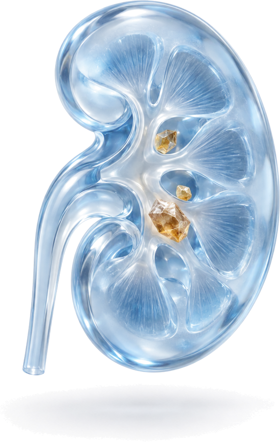
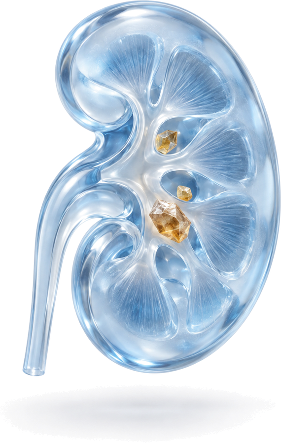
A kidney stone forms when substances in the urine, such as calcium, oxalate or uric acid, become concentrated and create crystals that gradually grow into a hard mass. Stones form in the kidney; once they move down into the tube that drains the kidney (the ureter), they are called ureteral stones. Because of its warm climate and dietary habits, Turkey is one of the countries where kidney stones are common.
The most common type is the calcium oxalate stone. Uric acid, calcium phosphate, infection (struvite) and cystine stones can also occur. Knowing the type of stone directly affects both treatment and advice on preventing new stones.
Symptoms of kidney stones
Stones sitting quietly in the kidney may cause no symptoms for a long time and may be found by chance on an ultrasound done for another reason. When a stone moves and blocks the urinary tract, the symptoms are usually sudden and obvious:
- Severe, wave-like pain spreading from the flank to the groin and genital area (renal colic)
- Nausea and vomiting
- Blood in the urine (visible or detected in a urine test)
- Frequent urination and a burning feeling when passing urine
- A frequent urge to go to the toilet as the stone approaches the bladder
When to seek urgent care: If stone pain is accompanied by fever and chills, if the amount of urine has dropped markedly, or if the pain cannot be controlled with painkillers, seek medical care without delay. A blocked and infected kidney needs urgent treatment.
How is it diagnosed?
During the consultation, the doctor asks when the symptoms started, whether you have passed stones before, which medications you take and whether there is a family history of stones. This is followed by:
- Urine test: Shows blood, infection and crystals.
- Blood tests: Assess kidney function and calcium and uric acid levels.
- Ultrasound: Involves no radiation; it is the first step in showing a stone in the kidney and swelling of the kidney (hydronephrosis).
- Non-contrast computed tomography (CT): Shows the size, location and density of the stone most accurately. Treatment is usually planned on the basis of this scan.
Kidney stone treatment options
The choice of treatment depends on the size, location and hardness of the stone, the anatomy of the kidney and the patient’s general health, all considered together. There is no single right method for every patient.
Monitoring and medication
Small kidney stones that cause no symptoms can be monitored with regular check-ups. For small stones that have moved into the ureter, plenty of fluids, painkillers and medication that relaxes the ureter may allow the stone to pass on its own. Uric acid stones can be dissolved with medication that makes the urine less acidic.
ESWL (shock wave lithotripsy from outside the body)
In ESWL, shock waves focused from outside the body break the stone into small fragments, which are then passed in the urine. It usually requires no anaesthesia and is done as an outpatient procedure. It is mostly used for kidney stones smaller than 2 cm that are not very hard. Some patients may need more than one session.
RIRS (incision-free laser kidney stone surgery)
In RIRS, a thin, flexible endoscope is passed through the urinary tract into the kidney, and the stone is broken up or turned to dust with laser energy. There is no incision in the skin. It is often preferred for stones that could not be broken with ESWL, stones in the lower part of the kidney and patients taking blood thinners. For details, see the RIRS page.
URS (ureteral stone surgery)
For stones that have moved into the ureter and do not pass on their own, a thin endoscope is passed through the urinary tract to reach the stone, which is then broken up with a laser and removed. No incision is needed.
PCNL (percutaneous nephrolithotomy)
For kidney stones larger than 2 cm and for staghorn stones that fill the inside of the kidney, the kidney is entered through a channel of about 1 cm made in the flank, and the stone is broken up and removed. In large stones, it is a method that achieves a high stone clearance rate in a single session. For details, see the PCNL page.
After treatment
After incision-free methods, most patients return to their daily lives within a short time. Mild blood in the urine and a burning sensation may occur in the first days; these usually settle on their own. For patients with a double-J stent, the date for stent removal is planned at discharge. The result of treatment is assessed with a follow-up scan a few weeks later.
Preventing kidney stones
Stone treatment does not end with removing the stone. To prevent recurrence:
- Drink about 2.5–3 litres of fluid spread over the day; aim for pale-coloured urine.
- Cut down on salt. Too much salt increases the calcium in the urine.
- Eat red meat and animal protein in moderation.
- Do not stop calcium on your own; calcium from food may reduce the risk of calcium oxalate stones.
- Have the passed or removed stone analysed; if you form stones frequently, ask for a metabolic evaluation.
Kidney stone treatment in Bursa, Turkey
Op. Dr. Mücahit Kabar graduated from Uludağ University Faculty of Medicine and completed his urology residency at the Urology Clinic of Ankara Training and Research Hospital. He has scientific studies on retrograde intrarenal surgery and percutaneous nephrolithotomy published in international peer-reviewed journals. He sees patients with kidney and ureteral stones in Bursa (Nilüfer) at Özel Medicabil Nilüfer Hastanesi (weekdays 08:30–12:00) and Özel Jimer Hastanesi (weekdays 13:00–17:00).
Consultations are held in English or Turkish. The hospitals’ international patient departments can arrange interpreters for other languages and help with airport transfers and accommodation. If you are travelling from abroad, you can book your appointment in advance through the online booking page or appointment line of Özel Medicabil Nilüfer Hastanesi (weekdays 08:30–12:00, +90 444 8 112) or Özel Jimer Hastanesi (weekdays 13:00–17:00, +90 444 45 67). You can share your CT or ultrasound reports beforehand via WhatsApp (+90 541 622 54 82). More information is available on the international patients page and the contact page.