What is RIRS?
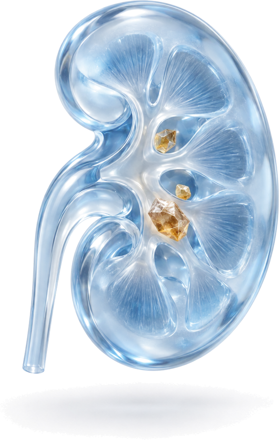
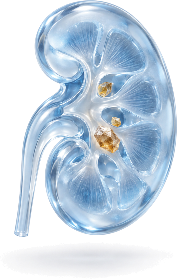
RIRS, short for retrograde intrarenal surgery, is an endoscopic method of treating kidney stones without any incision in the skin. “Retrograde” means that the kidney is reached against the direction of urine flow, that is, through the urethra, the bladder and the ureter.
The flexible ureterorenoscope used in the procedure is a very thin camera with a steerable tip. This allows the surgeon to reach the different chambers of the kidney (the calyces), even the hard-to-reach lower parts. Once the stone is seen, a thin laser fibre passed through the endoscope breaks it into small fragments or turns it to dust. This is why it is also known as laser kidney stone surgery.
When is RIRS used?
The choice of treatment is always based on the size, location and hardness of the stone and the patient’s general health, considered together. RIRS is often preferred in the following situations:
- Kidney stones smaller than 2 cm
- Stones that could not be broken with ESWL or that look hard on CT
- Stones in the lower part of the kidney (lower pole); fragments in this area may not pass easily after ESWL
- Patients taking blood thinners who cannot safely stop them
- Overweight (obese) patients, in whom it may be harder for shock waves from outside the body to reach the stone
- Selected patients with congenital differences in the position or structure of the kidney
- Treatment in the same session for patients with stones in both the kidney and the ureter
In suitable patients, RIRS can also be used for stones larger than 2 cm; in that case, the possibility of needing more than one session, and PCNL as an alternative, are discussed with the patient separately.
Preparing for surgery
Treatment is usually planned with a non-contrast CT scan, which shows the size, location and density of the stone and the anatomy of the kidney.
A urine test and urine culture are requested before surgery. Any urinary tract infection must be treated beforehand. Blood tests and an anaesthetic assessment are carried out. Always tell your doctor about all the medications you take, especially blood thinners.
In some patients, the ureter may be too narrow for the endoscope to pass safely. In that case, rather than forcing it, a double-J stent is placed first, and RIRS is carried out in a second session after a few weeks, once the ureter has widened.
How is RIRS performed?
- Under general or spinal anaesthesia, the bladder is entered first and a thin guide wire is placed in the ureter.
- If needed, a thin sheath (ureteral access sheath) is placed along the ureter to allow the endoscope to pass easily.
- The flexible ureterorenoscope is advanced into the kidney and the stone is located.
- The stone is broken up or turned to dust with laser energy. Suitably sized fragments can be removed with a fine basket catheter; dust-like fragments are passed in the urine.
- The kidney is checked and, if needed, a temporary double-J stent is placed to complete the procedure.
What is a double-J stent and why is it placed?
A double-J stent is a thin, soft tube with curled ends placed between the kidney and the bladder. It helps prevent swelling or small stone fragments from blocking the flow of urine after surgery. The stent is not visible from the outside.
A stent is not needed in every patient; the decision is made during surgery. Patients with a stent may experience frequent urination, urgency, mild blood in the urine and flank pain when passing urine. These complaints usually disappear once the stent is removed. The removal date is planned at discharge, and removing the stent on time is very important: forgotten stents can become encrusted with stone and infected.
Recovery
- Most patients go home the same day or the next day.
- Mild blood in the urine and a burning sensation may occur in the first days; with plenty of fluids, these usually settle quickly.
- Most people can return to desk work within a few days.
- The result of treatment is assessed with a follow-up scan a few weeks later. If significant stone fragments remain, an additional procedure may be needed.
What to watch for after going home
- Drink plenty of fluids spread over the day; aim for pale-coloured urine.
- Take the painkillers and other medications your doctor recommends regularly.
- Avoid heavy lifting and strenuous exercise in the first days; light walks are fine.
- Ask your doctor when to restart blood thinners.
- Do not miss your stent removal and follow-up appointments.
Advantages and limitations of RIRS
The main advantages of RIRS are that there is no incision in the skin and that the stone can be seen directly while it is broken up. This makes it an effective option for hard stones in which ESWL may be insufficient and for stones in hard-to-reach parts of the kidney. The low risk of bleeding means it can also be used in patients taking blood thinners. The hospital stay is short and the return to daily life is usually quick.
RIRS also has some limitations. It requires general or spinal anaesthesia. With very large stones, the procedure may take longer and more than one session may be needed. In patients with a stent, stent-related complaints may last from a few days to a few weeks. This is why the method is chosen together with the patient, after discussing their expectations.
Possible risks
RIRS requires no incision and is generally considered a safe method. Still, like any surgical procedure, it carries some risks:
- Blood in the urine (usually mild and temporary)
- Urinary tract infection and fever; rarely, hospital treatment may be needed
- Stent-related complaints
- Injury to the ureter; this is rare and usually heals with a stent
- The need for a second session because of remaining stone fragments
After discharge, seek medical care without delay if you have high fever, chills, unbearable pain or a marked drop in urine output.
RIRS, ESWL and PCNL compared
- ESWL: The stone is broken with shock waves from outside the body; anaesthesia is usually not needed. It may be less effective for hard stones, lower pole stones and overweight patients, and more than one session may be needed.
- RIRS: No incision; the stone is broken with a laser under direct vision. It requires anaesthesia, and a stent may be needed.
- PCNL: The kidney is entered through a small channel made in the flank. For stones larger than 2 cm and staghorn stones, it achieves high stone clearance in a single session. For details, see the PCNL page.
Which method is right for you is decided individually after examination and a CT scan. For general information, see the kidney stone treatment page.
RIRS in Bursa, Turkey
Op. Dr. Mücahit Kabar graduated from Uludağ University Faculty of Medicine and completed his urology residency at the Urology Clinic of Ankara Training and Research Hospital. He has scientific studies published in international peer-reviewed journals on the effectiveness of retrograde intrarenal surgery in obese patients and on RIRS in a crossed fused ectopic kidney. He sees patients with kidney stones in Bursa (Nilüfer) at Özel Medicabil Nilüfer Hastanesi (weekdays 08:30–12:00) and Özel Jimer Hastanesi (weekdays 13:00–17:00).
Consultations are held in English or Turkish. The hospitals’ international patient departments can arrange interpreters for other languages and help with airport transfers and accommodation. If you are travelling from abroad, you can book your appointment in advance through the online booking page or appointment line of Özel Medicabil Nilüfer Hastanesi (weekdays 08:30–12:00, +90 444 8 112) or Özel Jimer Hastanesi (weekdays 13:00–17:00, +90 444 45 67). You can share your CT reports beforehand via WhatsApp (+90 541 622 54 82). More information is available on the international patients page and the contact page.