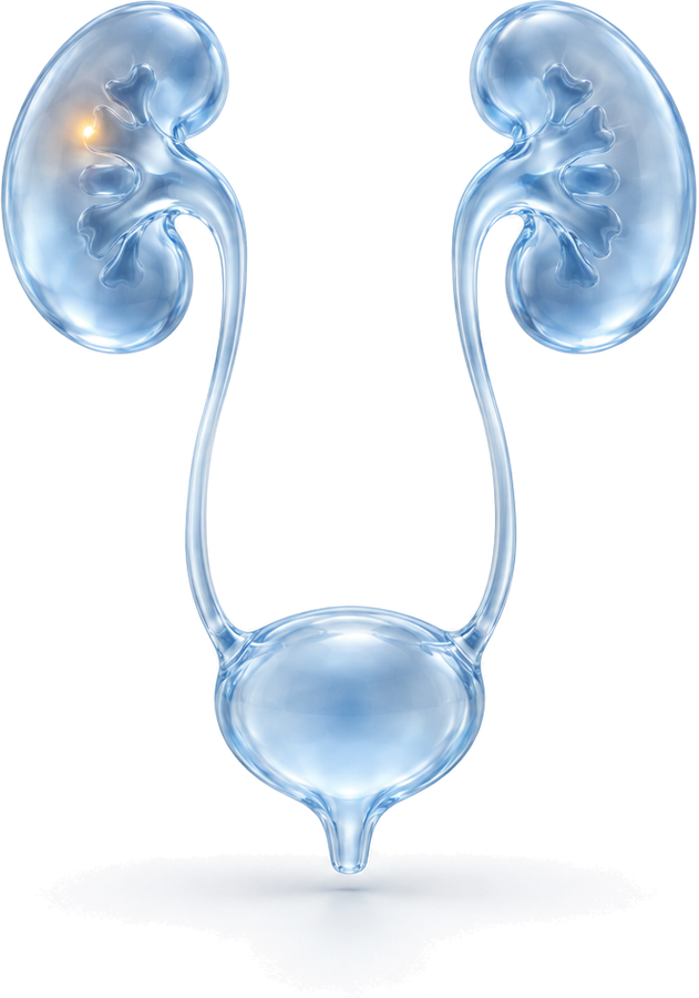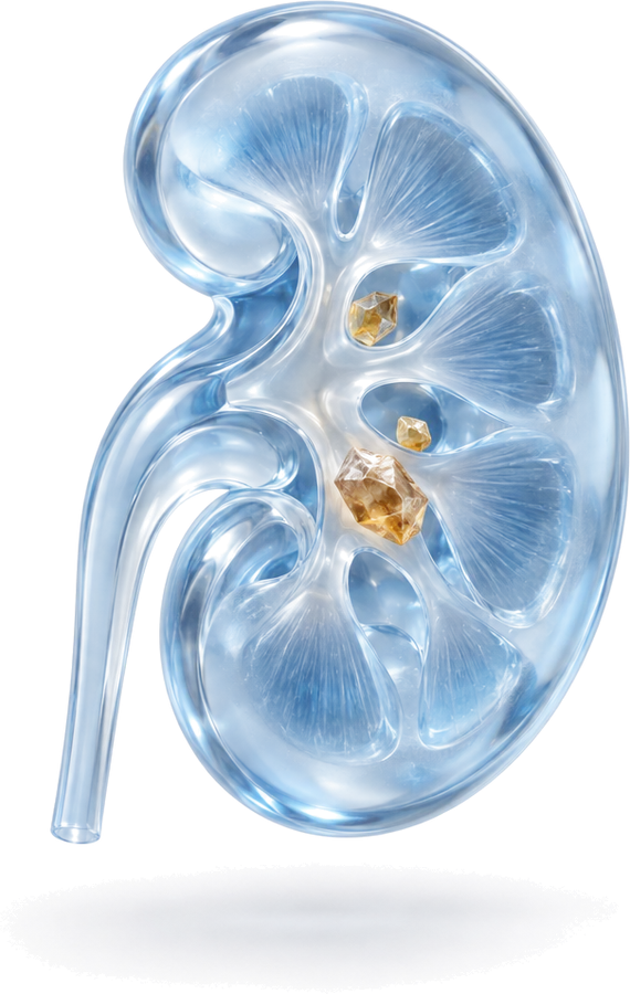
What is kidney cancer?
Kidney cancer is the general name for malignant tumours that develop from kidney tissue. The most common type in adults is renal cell carcinoma (RCC). Renal cell carcinoma also has subtypes: the most common is the clear cell type, followed by the papillary and chromophobe types. The subtype gives important information about how the tumour behaves and how follow-up should be planned. For example, the chromophobe type is less common and is generally considered to grow more slowly. The subtype, grade and stage of the tumour can only be confirmed by pathological examination of the tissue removed at surgery, and the follow-up plan after surgery is based on this information.
Today, thanks to the widespread use of ultrasound and CT, many kidney tumours are found by chance, while still small and without symptoms, on imaging done for another complaint. When tumours are caught early, kidney-sparing treatment options can be used more often.
Risk factors
- Smoking
- Being overweight and obesity
- High blood pressure
- Long-term dialysis and advanced chronic kidney disease
- Certain inherited conditions such as von Hippel-Lindau disease, and a family history of kidney cancer
Stopping smoking, maintaining a healthy weight and keeping blood pressure under control are among the main recommendations for reducing risk.
Symptoms
Kidney tumours are usually silent in the early stages. As the tumour grows, the following symptoms may occur:
- Blood in the urine
- Flank or back pain
- A mass that can be felt in the abdomen
- Unexplained weight loss, fatigue, fever or anaemia
None of these symptoms is specific to kidney cancer; however, blood in the urine in particular always requires a urological assessment.
How is it diagnosed?
Imaging
- Ultrasound: This is usually how a mass is first noticed. It helps distinguish a simple cyst from a solid mass.
- Contrast-enhanced computed tomography (CT): The key method for assessing kidney masses. The size and location of the mass, its relationship to the blood vessels and any possible spread are assessed with this scan.
- Magnetic resonance imaging (MRI): May be preferred for patients with a contrast allergy or impaired kidney function, and for assessing some complex cysts.
Cystic masses are assessed according to their imaging features using the Bosniak classification. Simple cysts need no follow-up or treatment, while some complex cysts may need monitoring or surgery.
Small renal masses and biopsy
Masses generally 4 cm or smaller are called small renal masses. Some of these are benign. A biopsy is not needed in every patient, but it can be done to guide the treatment decision in patients for whom surveillance or ablation is planned, or when imaging is inconclusive.
Not every kidney mass is cancer
Learning that a mass has been found in your kidney can be worrying, but not every mass is malignant. Simple cysts are very common and usually need no treatment at all. Solid masses also include benign tumours such as oncocytoma and angiomyolipoma. An angiomyolipoma can often be recognised on imaging because of the fat tissue it contains. Some benign tumours such as oncocytoma, however, cannot be distinguished from kidney cancer on imaging, and the final diagnosis can only be made by pathological examination of the removed tissue.
For this reason, in every patient with a kidney mass, the size of the mass, its imaging features, its growth rate and the patient’s general health are assessed together. The aim is to treat malignant tumours in good time while also avoiding unnecessary loss of a kidney. This is one of the reasons why kidney-sparing surgery is preferred in suitable patients.
Kidney cancer treatment
Treatment is planned individually, taking into account the size of the tumour, its location in the kidney, its stage, the condition of the other kidney and the patient’s general health.
Partial nephrectomy (kidney-sparing surgery)
For small tumours confined to the kidney, in technically suitable patients, only the tumour is removed with a margin of healthy tissue and the rest of the kidney is preserved. This approach helps preserve kidney function in the long term and is recommended in current guidelines as the preferred option for suitable patients.
Radical nephrectomy
If the tumour is large, located in the centre of the kidney or has grown into the blood vessels, or if partial nephrectomy is not technically suitable, the whole kidney is removed together with the surrounding fat tissue. People whose other kidney is healthy can continue to lead a normal life with one kidney.
Laparoscopic approach
Both partial and radical nephrectomy can be performed in suitable patients using a laparoscopic technique, that is, through a few small openings guided by a camera. Compared with open surgery, there is generally less pain after the operation and a shorter hospital stay and recovery. For very large tumours or those that have grown into surrounding tissues, open surgery may be more appropriate.
Active surveillance and ablation
In selected patients with a small mass who are older or have a high surgical risk because of other illnesses:
- Active surveillance: The mass is imaged at regular intervals; if it grows, the treatment plan is reassessed.
- Thermal ablation: The tumour is destroyed by freezing (cryoablation) or with heat energy (radiofrequency or microwave) through a needle placed through the skin.
Advanced disease
When the disease has spread beyond the kidney, treatment is planned together with medical oncology. Immunotherapy and targeted therapies are the main options used in this group; in selected patients, surgery may also be part of the treatment plan.
After surgery and risks
After laparoscopic surgery, patients usually go home within a few days and return to daily life within a few weeks. As with any surgery, there are risks such as bleeding, infection and injury to nearby organs; after partial nephrectomy, urine leakage may also occur. These risks are discussed in detail before surgery.
Follow-up
After kidney tumour surgery, imaging and blood tests are carried out at intervals set according to the pathology result and risk group. Follow-up aims to detect any recurrence early and to monitor the function of the remaining kidney tissue.
To protect the kidneys after surgery, it is recommended to keep blood pressure under control, drink enough fluids, avoid long-term use of painkillers that can harm the kidneys without medical advice, and stop smoking. If you notice blood in the urine, flank pain or unexplained weight loss between check-ups, see your doctor without waiting.
Kidney tumour treatment in Bursa, Turkey
Op. Dr. Mücahit Kabar graduated from Uludağ University Faculty of Medicine and completed his urology residency at the Urology Clinic of Ankara Training and Research Hospital. He has scientific studies evaluating chromophobe renal cell carcinoma in a radical nephrectomy series and examining benign lesions removed by nephrectomy for a suspected renal mass. He sees patients with kidney masses in Bursa (Nilüfer) at Özel Medicabil Nilüfer Hastanesi (weekdays 08:30–12:00) and Özel Jimer Hastanesi (weekdays 13:00–17:00).
Consultations are held in English or Turkish. The hospitals’ international patient departments can arrange interpreters for other languages and help with airport transfers and accommodation. If you are travelling from abroad, you can book your appointment in advance through the online booking page or appointment line of Özel Medicabil Nilüfer Hastanesi (weekdays 08:30–12:00, +90 444 8 112) or Özel Jimer Hastanesi (weekdays 13:00–17:00, +90 444 45 67). You can share your CT or MRI reports beforehand via WhatsApp (+90 541 622 54 82). More information is available on the international patients page and the contact page.